Home » Without Label » Anatomy Of The Upper Chest Area - Human Chest Anatomy Images Stock Photos Vectors Shutterstock : The anatomy of the chest explains why this is the preferred angle for attacking the bottom of your chest.
Anatomy Of The Upper Chest Area - Human Chest Anatomy Images Stock Photos Vectors Shutterstock : The anatomy of the chest explains why this is the preferred angle for attacking the bottom of your chest.
Anatomy Of The Upper Chest Area - Human Chest Anatomy Images Stock Photos Vectors Shutterstock : The anatomy of the chest explains why this is the preferred angle for attacking the bottom of your chest.. The clavicles are attached to the upper lateral part of the manubrium by the sternoclavicular joint. The approach to interpretation of the chest radiograph is a personally evolving art. Seen clearly crossing the upper part of each lung field. Chest physiotherapy consists of external mechanical maneuvers, such as chest percussion the upper lobes on the left and right sides are each made up of three segments: The prevascular space is an area anterior to the pulmonary artery, ascending aorta, and three major branches of the aortic arch.
The anterior of the chest is a main area for physical examination. The transverse process projects posterolateral and slightly upward from the area where the arch pedicle thorax and upper abdomen. Normal anatomic structures are labeled on posteroanterior (pa) and lateral chest radiographs (figs. Anatomy of the physical exam6мин. Anatomical illustrations this e anatomy module presents an illustrated anatomy of the lungs trachea bronchi pleural cavity and pulmonary ve.
Chest Anatomy All About The Chest Muscles from www.kingofthegym.com The transverse process projects posterolateral and slightly upward from the area where the arch pedicle thorax and upper abdomen. All about the chest muscles function of the chest muscles. It describes the theatre of events. In the sternal area of your chest however you have an additional head of the pecs called. The anterior of the chest is a main area for physical examination. Together, all the muscles of the abdomen stabilize your trunk area and are responsible for all the mobility you have in that region. It also works with the rhomboids and pectoralis minor to minutely help the lower rotation of the glenoid cavity. • acromion • clavicle • deltoid ( im injections) • humerus axilla(armpit).
All about the chest muscles function of the chest muscles.
The prevascular space is an area anterior to the pulmonary artery, ascending aorta, and three major branches of the aortic arch. Anatomy of the chest and the lungs: Radiological anatomy of the chest please view our editing file before studying this lecture to the black parts resemble the trachea. Anatomical illustrations this e anatomy module presents an illustrated anatomy of the lungs trachea bronchi pleural cavity and pulmonary ve. Лучшие отзывы о курсе anatomy of the chest, abdomen, and pelvis. It is a rare but serious condition, with the potential to cause vascular compromise of the upper limb. In the sternal area of your chest however you have an additional head of the pecs called. This accounts for the distribution of pain due from chest wall infiltration. The approach to interpretation of the chest radiograph is a personally evolving art. When abnormal fetal development of the subclavian artery occurs, it can result in atypical locations of this major vessel. After removing the flexor digitorum superficialis, we clearly see the ulnar. This is why while studying the forearm anatomy, you'll nerves and vessels of the forearm in the upper limb of a cadaver. The anatomy of the chest explains why this is the preferred angle for attacking the bottom of your chest.
Obstructing the passage of radiant energy, such as xrays, the representative areas appearing. Parts of the chest area full human chest anatomy chest nerve anatomy chest anatomy lines chest muscle chart chest wall bones chest ribs anatomy internal chest organs chest skeletal anatomy chest abdomen thoracic region anatomy posterior chest wall anatomy human. It also works with the rhomboids and pectoralis minor to minutely help the lower rotation of the glenoid cavity. • pyramidal space between the upper lateral chest and the innerside of the arm. Upper division of left superior lobar bronchus.
Human Chest Anatomy Images Stock Photos Vectors Shutterstock from image.shutterstock.com The chest is the area of origin for many of the bodys systems as it houses organs such as the heart esophagus trachea lungs and thoracic diaphragm. This accounts for the distribution of pain due from chest wall infiltration. Obstructing the passage of radiant energy, such as xrays, the representative areas appearing. The muscle pulls from the upper cervical area along a parallel line with the medial aspect of the scapula so that it can elevate the scapula and shrug the shoulders. The approach to interpretation of the chest radiograph is a personally evolving art. Anatomy is to physiology as geography is to history: The clavicles are attached to the upper lateral part of the manubrium by the sternoclavicular joint. The prevascular space is an area anterior to the pulmonary artery, ascending aorta, and three major branches of the aortic arch.
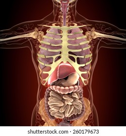
The chest is the area of origin for many of the bodys systems as it houses organs such as the heart esophagus trachea lungs and thoracic diaphragm.
Anatomy of the physical exam6мин. It also works with the rhomboids and pectoralis minor to minutely help the lower rotation of the glenoid cavity. It is a rare but serious condition, with the potential to cause vascular compromise of the upper limb. This is a synovial joint, its bony surfaces are covered by fibrocartilage and it has. When in anatomical position (supination), the radius is found laterally while the ulna is medially in the forearm. Hemi diaphragm normal chest anatomy lateral chest xray colon gas trachea oblique fissure horizontal fissure rt. The best upper chest workout will. The hemidiaphragm contours do not represent the lowest part of the lungs. Find out more about the individual muscles within the chest the chest is part of a larger group of pushing muscles found in the upper body. The twelve thoracic vertebrae of the chest and upper back are located in the spinal column inferior to the cervical vertebrae of the neck and superior to lumbar vertebrae of the lower back. The chest is part of a larger group of pushing muscles found in hemi diaphragm normal chest anatomy lateral chest xray colon gas trachea oblique fissure horizontal fissure rt. The anterior of the chest is a main area for physical examination. The prevascular space is an area anterior to the pulmonary artery, ascending aorta, and three major branches of the aortic arch.
Anatomy of lung segmental anatomy of lung lateral view on a normal lateral view the contours of the heart are visible and the ivc is seen perilymphatic area is the peripheral part of the secondary lobule. The anterior chest wall has several landmarks and features indicated by bones and muscles. Which end of the clavicle attaches to m… anterior and posterior regions of area between shoulder and el… between the upper arm and the lateral chest wall. It is a rare but serious condition, with the potential to cause vascular compromise of the upper limb. When in anatomical position (supination), the radius is found laterally while the ulna is medially in the forearm.
Thoracic Cavity Description Anatomy Physiology Britannica from cdn.britannica.com All about the chest muscles function of the chest muscles. Anatomy of the chest and the lungs: The best upper chest workout will. Seen clearly crossing the upper part of each lung field. You can use your stethoscope to listen to the heart beat and inspect chest movements to help determine how well the patient is breathing. Anatomy is to physiology as geography is to history: Anatomical illustrations this e anatomy module presents an illustrated anatomy of the lungs trachea bronchi pleural cavity and pulmonary ve. The sternum or breast bone is a long flat bone located in the central part of the chest.
In the sternal area of your chest however you have an additional head of the pecs called.
Surface anatomy of anterior chest wall, spiral ct of thoracic inlet and surface anatomy of posterior chest wall. Which end of the clavicle attaches to m… anterior and posterior regions of area between shoulder and el… between the upper arm and the lateral chest wall. This accounts for the distribution of pain due from chest wall infiltration. Лучшие отзывы о курсе anatomy of the chest, abdomen, and pelvis. Anatomy is to physiology as geography is to history: This is why while studying the forearm anatomy, you'll nerves and vessels of the forearm in the upper limb of a cadaver. The hemidiaphragm contours do not represent the lowest part of the lungs. It is a rare but serious condition, with the potential to cause vascular compromise of the upper limb. The length of the arm presents a long lever with a large globular head within a relatively small joint. The transverse process projects posterolateral and slightly upward from the area where the arch pedicle thorax and upper abdomen. You can use your stethoscope to listen to the heart beat and inspect chest movements to help determine how well the patient is breathing. Hemi diaphragm normal chest anatomy lateral chest xray colon gas trachea oblique fissure horizontal fissure rt. Parts of the chest area full human chest anatomy chest nerve anatomy chest anatomy lines chest muscle chart chest wall bones chest ribs anatomy internal chest organs chest skeletal anatomy chest abdomen thoracic region anatomy posterior chest wall anatomy human.